Estimated reading time: 4 minutes
Back in 2020, we published a whitepaper about the need for updated data storage infrastructures to support advanced image generators in life sciences. We predicted that one of the key factors driving these new demands would be the growing use of artificial intelligence (AI) in high-end microscopy workflows.
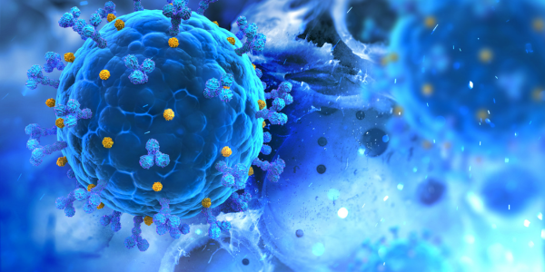
Now in 2023, we’re seeing that prediction come true across cryogenic electron microscopy (cryo-EM) and cryo-electron tomography (cryo-ET), scanning electron microscopy (SEM), and transmission electron microscopy (TEM). In other words, we’re in the early stages of an era of rapid progress in AI-enabled data analysis that will vastly expand the scope of these techniques.
As AI continues to evolve, we can expect these imaging generators to unveil new horizons, catalyzing breakthroughs across disciplines that will redefine the boundaries of knowledge. The future promises an era of AI-driven exploration, discovery, and innovation at the nanoscale. Let’s take a look.
Cryo-EM, Cryo-ET, SEM, and TEM at a Glance
Before we dive in, here’s a quick overview of how these high-end microscopy workflows compare.
| Technique | Purpose | Sample Preparation | Imaging Depth | Resolution | Application Areas |
| Cryo-EM | Visualize biological structures at near-atomic resolution | Rapid freezing and preservation in vitreous ice | 2D (single particles) or 3D (cryo-ET) | Near-atomic (~1-3 Å) | Structural biology, protein complexes, viruses |
| Cryo-ET | 3D visualization of cellular structures | Rapid freezing and preservation in vitreous ice | 3D | Nanoscale (few nanometers) | Cell organelles, cellular architecture, viruses |
| SEM | Surface morphology imaging | Coating with conductive layer or cryogenic preparation | 2D | Nanoscale (few nanometers) | Materials science, nanotechnology, biology |
| TEM | Internal structure imaging | Ultrathin sectioning and staining or cryogenic preparation | 2D | Atomic to nanoscale (sub-Å) | Materials science, biology, nanotechnology |
Both Cryo-EM and Cryo-ET excel in providing detailed insights into the internal structures of biological specimens, with Cryo-ET offering a 3D perspective. SEM, on the other hand, focuses on surface morphology, making it suitable for examining various materials’ outer features and particularly useful in nanotechnology and various industrial applications. TEM goes deeper, imaging internal structures with high resolution. Together, all these techniques let researchers unlock keys to advancements in medicine, materials, and beyond.
The Growing Use of AI in High-end Microscopy
As we all know by now, the time for suspecting that AI’s been overhyped has long passed. The reality is, it’s quickly becoming an essential tool that tackles the hurdles brought about by the massive data produced by these state-of-the-art microscopy methods.
AI-Powered Enhancement of Cryo-EM and Cryo-ET
As these microscopes become more sophisticated and continue to generate larger and more complex datasets, traditional data processing strategies in cryo-EM and cryo-ET are quickly becoming too costly in both user time and computational resources; this is where AI can change the game.
It’s true that AI efforts have been somewhat slow to take hold in this space owing to several factors, including historically poor data management, varying data types, and collaboration barriers. Another obstacle involves the lack of complete datasets needed for training the algorithms – as of right now, only about half of the maps in the Electron Microscopy Data Bank (EMDB) have a complete atomic structure.
But as activity in these areas surges, this is all quickly changing. With recent advances and better data management practices, several fully automated deep learning-based image processing approaches are currently being applied cryo-EM and cryo-ET workflows, including steps such as particle picking (e.g., DeepPicker, DeepEM, TOPAZ), 3D classification (e.g., CryoGAN, 3DFlex), resolution determination, map sharpening, and post processing (e.g., DeepRes).
AI’s reach into SEM and TEM
We’re seeing AI applications being used to accelerate time to insights and improve results in SEM and TEM as well. In a recent collaboration between POSTECH and the Korea Institute of Materials Science (KIMS), researchers used deep learning to develop a super-resolution imaging technique for SEM workflows. The outcome was nothing short of remarkable: microstructure image resolution was enhanced by factors of 4, 8, and even 16 times, leading to an exponential reduction in imaging time—up to 256 times faster compared to conventional SEM systems.
TEM, which is indispensable for understanding the internal arrangements of cells, nanoparticles, and even atomic structures, also stands to make great strides with AI-driven automation. AI-enabled microscopy data analytics, including machine learning enhanced tools for retrieving useful information from massive TEM imaging, diffraction, and spectroscopy datasets, are expected to radically accelerate workflows.
AI Makes More Possible in Less Time
So: What’s the big deal? The incorporation of AI in cryo-EM, cryo-ET, SEM, and TEM workflows is ultimately a pivotal step toward maximizing their potential.
- Speed and Efficiency: The automation enabled by AI significantly speeds up traditionally labor- and time-intensive processes like particle picking and map reconstruction, allowing researchers to focus on interpreting results rather than wrestling with data manipulation.
- Quality and Accuracy: AI-driven algorithms excel at tasks that require a high degree of precision, such as image denoising and segmentation. This enhances the quality and accuracy of reconstructed structures, providing researchers with more reliable data for their analyses.
- Overcoming Complexity: The complexity of biological systems and cellular structures demands sophisticated analysis techniques. AI’s ability to identify patterns and relationships within these intricate datasets enables researchers to uncover hidden insights that might have otherwise remained unknown.
- Democratization of Expertise: AI automates complex data processing tasks that traditionally required specialized expertise. This democratization of expertise allows researchers from diverse backgrounds to engage with high-end microscopy techniques without being held back by steep learning curves.
- Exploration of Uncharted Territories: The speed and efficiency that AI offers open the doors to exploring new frontiers in research. Researchers can now undertake larger and more ambitious projects, pushing the boundaries of our understanding in areas such as drug discovery, materials science, and neuroscience.
Data Storage and Management Solutions That Keep Pace
In this rapidly evolving landscape of AI-powered microscopy, having a solid foundation for data storage and management is crucial. As the volume of data generated by these cutting-edge techniques surges, Panasas is ready to provide the seamless infrastructure you need to keep the wheels of discovery turning.
Our storage solutions are designed to handle the data deluge coming from AI-boosted microscopy workflows. Whether it’s the extensive datasets from cryo-EM and cryo-ET or the lightning-fast results from SEM and TEM workflows, we ensure that your data is safely stored, easily accessible, and ready for analysis. With our reliable architecture and supremely simple management interface, your team can focus on science while we take care of the data. For more information on that front, head over to our microscopy solution brief here.